Were we successful?
The following patient was in the EP lab for a paroxysmal AF ablation.
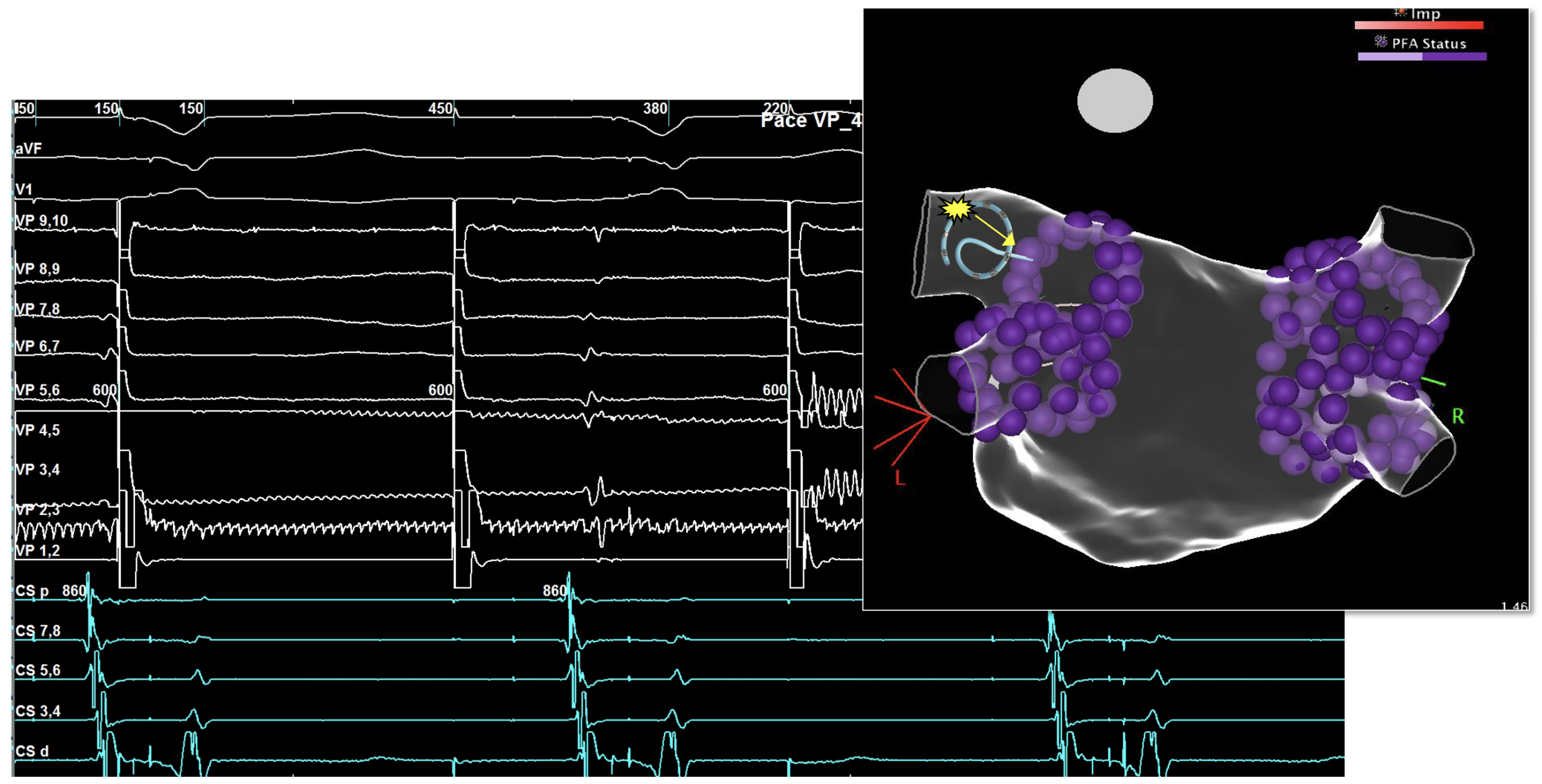
Pulsed Field Ablation (PFA) was utilized, but first, high-density mapping was performed of the left atrium.
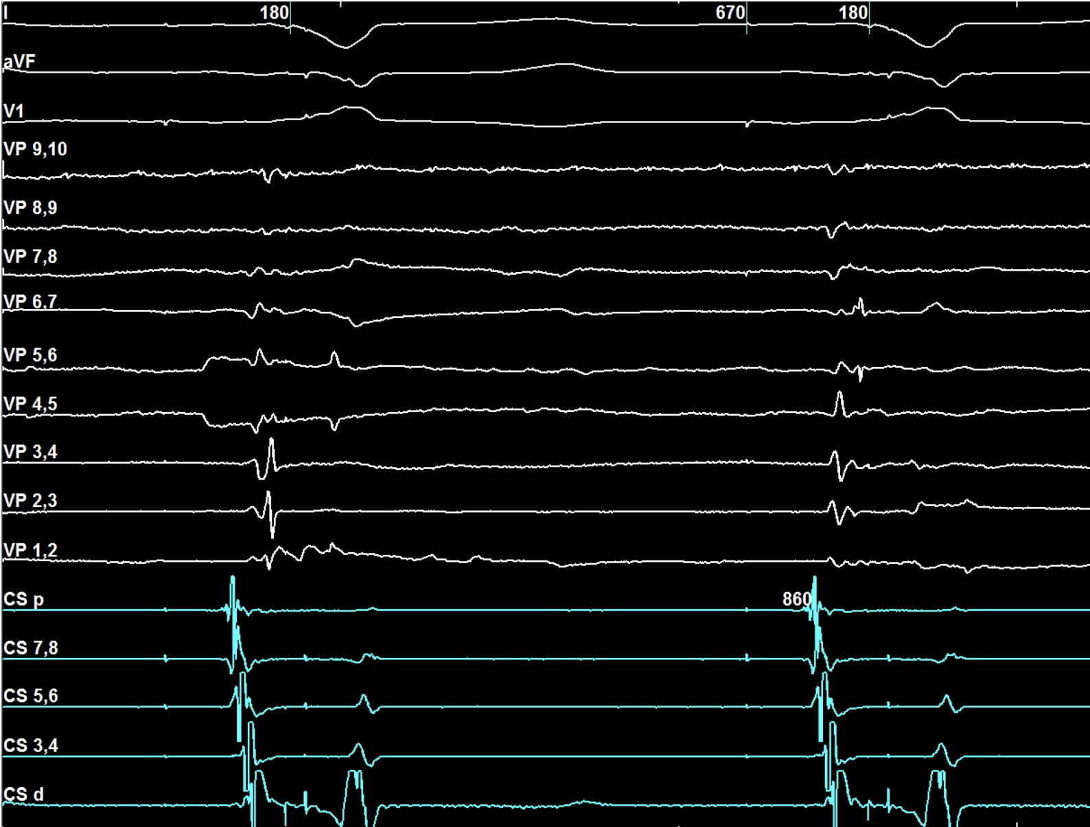
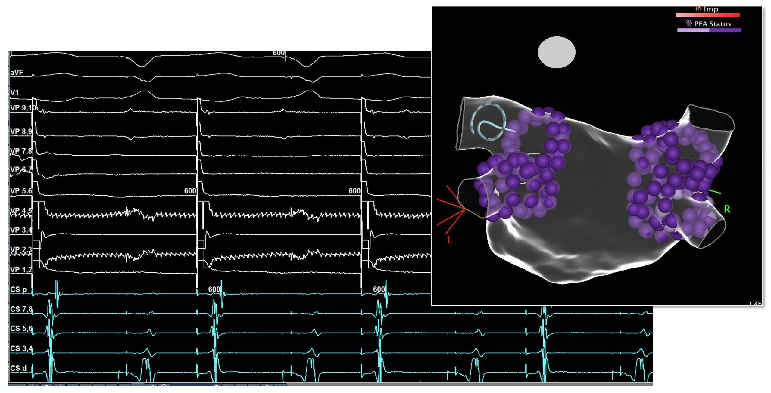
After completion of the lesion set, prior to remapping, pacing was attempted from the PFA catheter from each of the four veins. The electrogram displayed was recorded in the LSPV. Due to the proximity of the left atrial appendage, pacing was performed on each channel to differentiate the signals observed from the LAA.
What do you observe with PFA catheter pacing?
Answer:
We do not have exit block as the impulse can “exit” the pulmonary vein and enter the left atrium as seen by the CS activation immediately following the pacing in the LSPV. Further ablation is needed in this vein.
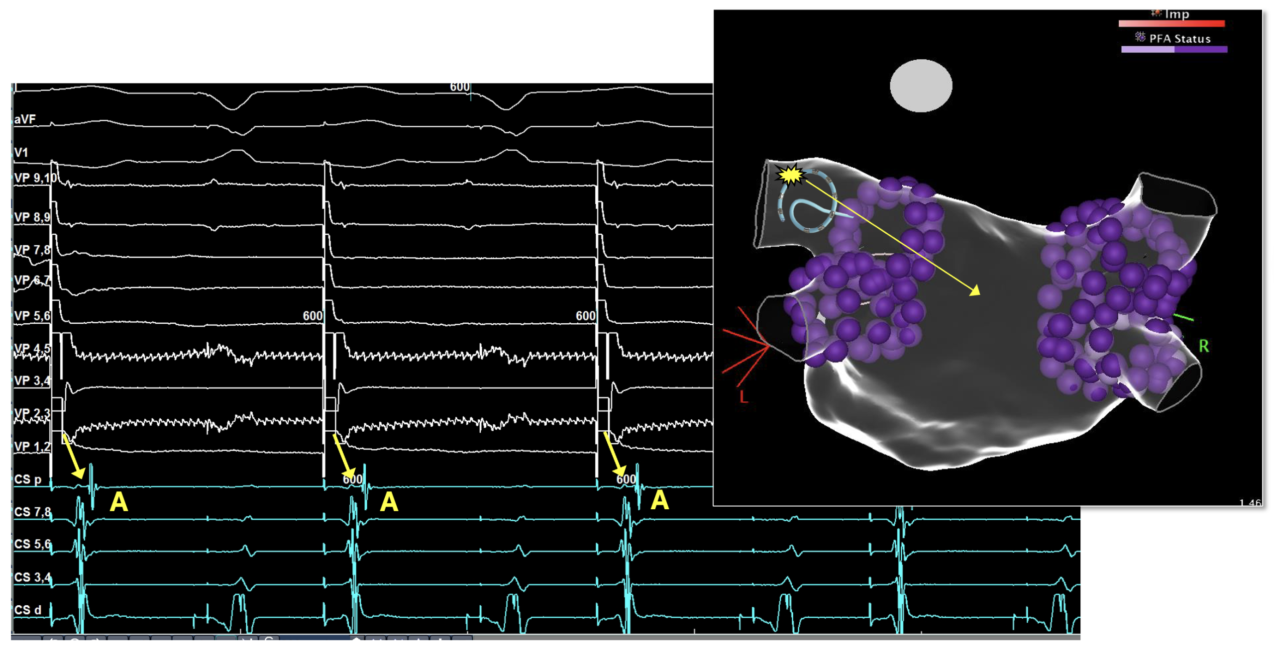
Now, after additional ablation was performed, what is observed?
Answer:
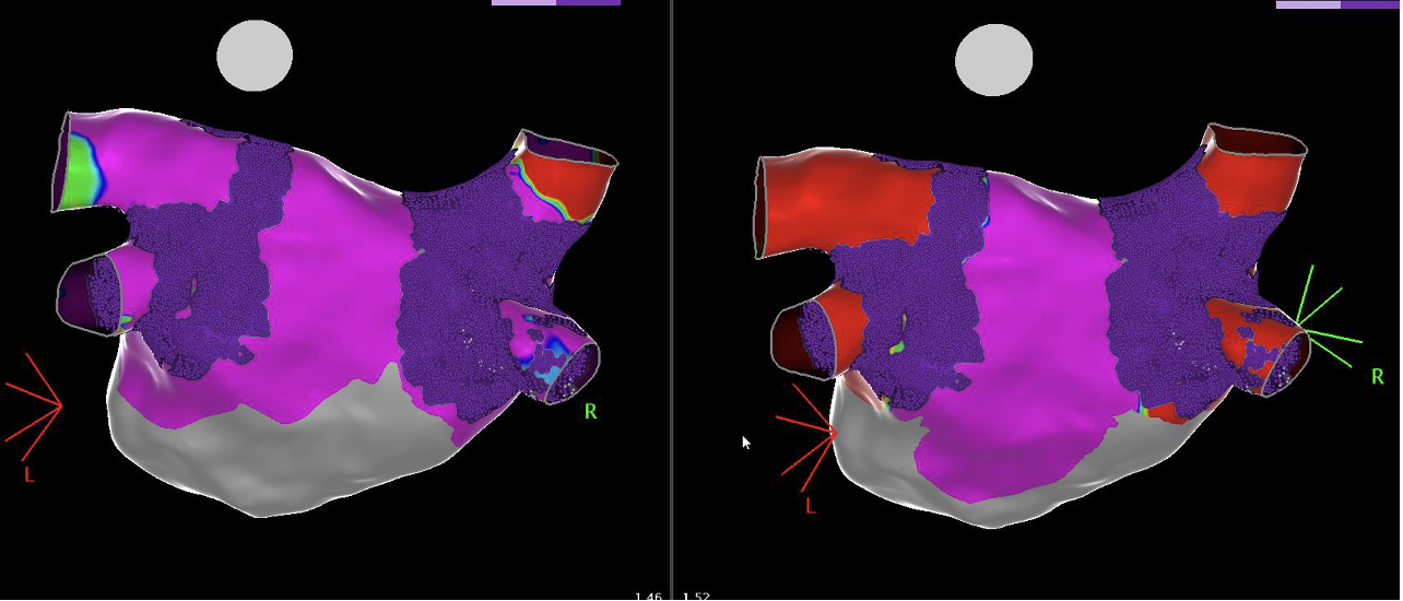
Now we can see that the pacing within the LSPV is no longer able to exit the vein and enter the left atrium. This is an example of exit block. No further ablation is needed; however, for completeness, the mapping catheter was reinserted for post-ablation substrate mapping. The image on the left is a PA view before PFA delivery (purple is considered healthy), the image on the right is after PFA delivery and shows low tissue voltage in the four pulmonary veins; no further ablation is needed. Notice the dark purple on each image is the PFA lesion set; there are various ways to display the lesions depending on the physician’s preference.