
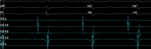
What is the catheter location in the following electrogram?
Answer
Near the Bundle of His
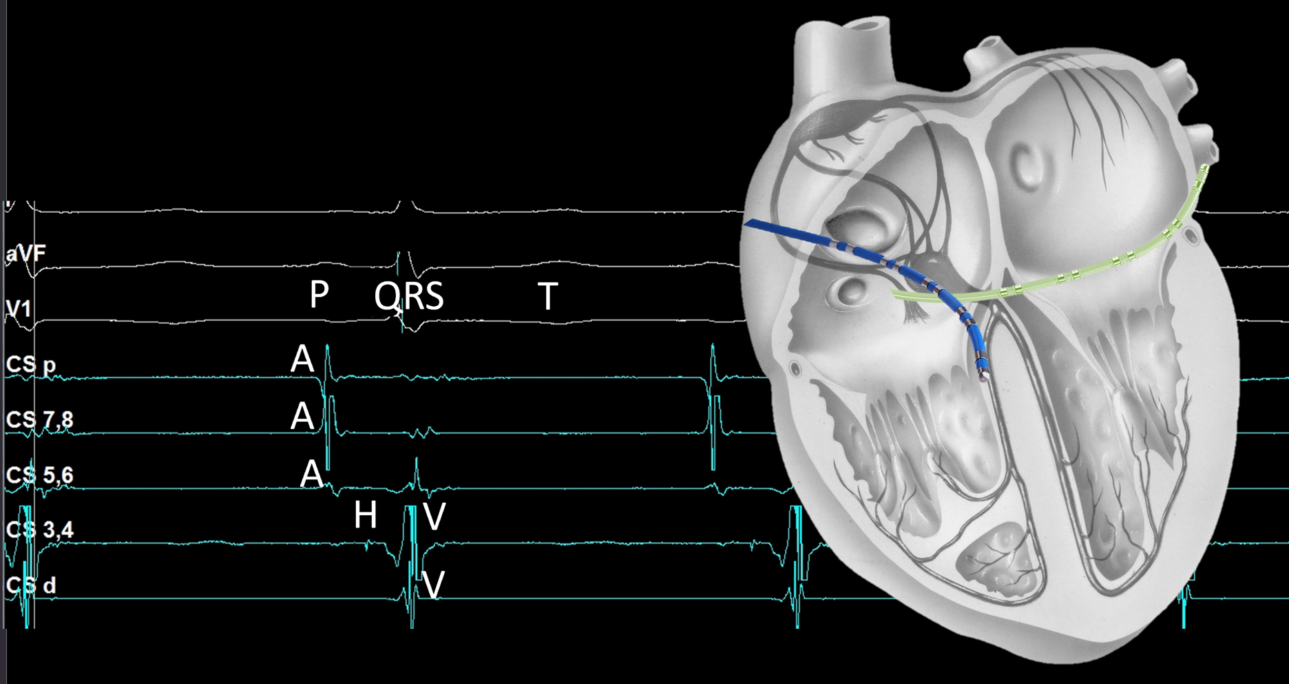
The catheter is labeled coronary sinus (CS); however, the signals do not reflect that location. Notice the A, H, and V signals recorded. Instead of placement in the coronary sinus (green catheter in the image), the catheter was placed near the bundle of His (blue catheter).
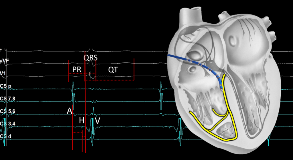
A physician may choose not to insert all four standard EP catheters to cutdown on cost as well as access sites if it is not needed. Therefore, do not rely on the labels but learn to recognize placement by the waveforms. This physician was recording the baseline measurements before properly placing the catheter in the CS. When recording the His measurements, we like to observe an A, H, and V all on the same channel. That is not the case in this example and the HV was measured as 18 ms. Recall, the HV is normally 35-55ms, so while a short HV may lead you to believe that there is an accessory pathway. In this example though, the catheter was simply advanced to far and is recording the right bundle potential instead…. Resulting in the short measurement. The catheter should be pulled back slightly to obtain a true His recording.
Please subscribe for a weekly link to the newest blog post.
For more information: EP Essentials – Understanding EP: A Comprehensive Approach


